- MR Angiography
- Perfusion imaging (ASL MRI)
- Edema imaging (T2, ADC)
- pH imaging (CEST)
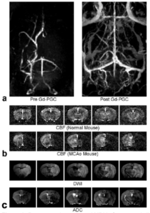
Figure 1: Representative structural MRI in five coronal slices of a transient MCAO mouse stroke model: (a) angiograms (maximum intensity projection) before and after the administration of Gd-PGC, (b) cerebral blood flow (CBF) maps acquired using ASL for normal and MCAO mice, (c) DWI and ADC maps (2 hrs after the onset of MCAO).